
 Tiếng Việt
 Indonesia
Causes:
The causative agent of subcutaneous viral infection and necrosis is similar to Parvovirus, its nuclear acid structure is DNA, and its diameter is 22 nm. Viruses parasitic in the nucleus of cells of the antenna gland, lymphatic system cells, gill cells, nerve cells, cannot hide (occlusion body) but can be included (inclusion body), they cause necrosis and swelling main character.
Syndromes:
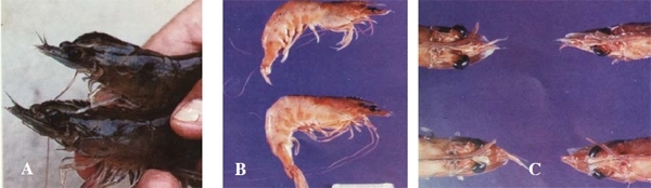
– Shrimp infected with IHHNV are often lethargic, weak, and deformed. Infected Black tiger shrimp (P.monodon), when dying, they often turns green, the abdominal muscles are cloudy. Vannamei shrimp (P. vannamei) exhibits stunted dysmorphic syndrome, juvenile shrimp (Juvenil) deformed main body, twisted antennae, rough or deformed chitinous shell.
The stunting coefficient in the white leg shrimp seed stock with IHHNV is usually from 10-30%, when the disease is severe, the stunting coefficient is as high as 30%, sometimes up to 50%. Shrimp P. stylirostris has an acute form of disease, very high mortality, the virus is transmitted from mother to larva (vertical) but does not show disease, often postlarvae 35 signs of disease observed are high mortality, virus Horizontal spread in postlarvae is very intense, adult shrimp sometimes show signs of disease or death.
– Histopathological examination of antennae gland cells, nerve cells and gill cells of shrimp infected with IHHNV, which may be embedded in the cell nucleus. In the early stage, it is usually small in the center of the nucleus, then it grows near the nucleus (Eosin’s color is red to dark red). The inclusion body contains many viruses.
Distribution:
IHNNV disease was detected in the US in the white leg shrimp (Penaeus vannamei), also known as the syndrome of stunting of South American white shrimp. The disease occurs from postlarvae to adult shrimp. The mortality rate of shrimp P. stylirostris is very high. The disease occurs in Singapore, Philippines, Thailand, Indonesia, and Malaysia.
IHHNV disease spreads both vertically and horizontally, the virus can be transmitted from broodstock to larval shrimp or infect early shrimp larvae.
In Vietnam, through histopathological analysis of the hepatopancreas of black tiger shrimp P.monodon Minh Hai, Soc Trang showed inclusions in the nucleus of the antennal cells of black tiger shrimp (Bui Quang Te, 1994) but the virus infection rate was low. Black tiger shrimp and vannamei shrimp (P. vannamei) cultured in Quang Ninh were diagnosed by PCR test and showed that shrimp were infected with IHHNV, cultured shrimps grew slowly and unevenly, the rate of stunted shrimp was 20-50% (Bui Quang Te, 2004).